Many folks we see daily have—in addition to blepharitis—rosacea, auto-immune disease, diabetes, allergy, contact lens use, history of ocular surgery, and glaucoma. This myriad of confounding chronic conditions, symptoms, and risk factors often have one common clinical finding: eyelid inflammation.
Some clinical studies suggest that 12–47% of patients with symptoms of foreign body sensation, irritation, itching, vision fluctuation, redness, stinging, and dryness have blepharitis. In my clinic, it runs closer to 70–80%.
The eyelids, including the meibomian glands, form the most important barrier against pathogens on the ocular surface. Treating eyelid inflammation early and regularly with effective therapy makes a profound impact on quality of life for many of our patients.
Thanks to instruments introduced in the last 10 years, we have more effective in-office treatments. Our current challenge is how best to keep the eyelid margins clean and the meibomian glands unclogged over the long term. In the past, we have sent patients home with instructions to clean their lids and apply warm compresses daily. Compliance has been low, and the conditions tend to reappear.
Addressing blepharitis on a continuum
We often characterize blepharitis by referring to anterior and posterior anatomical landmarks. Anterior involvement occurs mostly along the base of the lashes, eyelash follicles, and the cutaneous side of the eyelid. Posterior involvement typically refers to chronic inflammation that leads to meibomian gland disease. Patients may present with both conditions. Additionally, many patients with chronic staphylococcal and seborrheic blepharitis develop meibomian gland dysfunction (MGD).
Rather than segmenting anterior and posterior, I think of these anatomical landmarks as one important line of defense. Together, they are crucial in preventing ocular surface disease.
MGD presenting as terminal ductal obstruction and tissue atrophy leads to qualitative and quantitative changes in glandular secretions. Such abnormalities are evidence that a critical layer of defense is compromised.
Untreated MGD depletes normal meibum delivery to the ocular surface, leading to increased bacterial growth and early tear film evaporation.
To avoid vision-threatening corneal complications, it is important to understand the relationship between ocular surface compromise and chronic eyelid inflammation.
Biofilm’s role in dry-eye disease
Staphylococcal epidermitis and Staphylococcal aureus produce the protein adhesin, which gives biofilm its most important feature: sticking power. Adhesin ensures that bacteria adhere to the eyelid surface. The normal eyelid flora become over-colonized and induce pathologic inflammation over a patient’s lifetime.
The multi-laminar organization of this substrate provides a friendly environment for diverse bacteria living together in one complex community. This highly organized and protected community continues to increase within the biofilm with age, eventually becoming vastly over-colonized.
Biofilm thickens over time, resulting in production of pro-inflammatory factors. This is especially evident in contact-lens wearers. This population struggles with more dry-eye disease, MGD, and blepharitis largely due to the contact lens being a friendly surface for biofilm adhesion.
Over-colonization and overgrowth in chronic blepharitis lead to enhanced cell-mediated immunity involving staphylococcal antigens, which attach to the bacterial antigen-binding receptors on the corneal epithelium, provoking inflammation.
Staphylococcal marginal keratitis (MK) is an immune-mediated response to the bacterial antigens present in lid margin disease. MK typically presents with peripheral corneal infiltrates with an epithelial defect adjacent to the limbus. The infiltrate stains with fluorescein and is separated from the limbus by a clear cornea. Patients with marginal keratitis may present with bilateral disease and are usually uncomfortable.
New approach to long-term treatment
When treating acute staphylococcal MK, the most important consideration is to not neglect underlying chronic conditions. These patients need a long-term plan so that acute episodes of MK are less likely.
I first reduce the local population of staphylococci, improve MGD, and reduce the local immune response using a combination of antibiotic (erythromycin in children) and anti-inflammatory drops.
Debriding eyelid margins and removing pro-inflammatory elements is arguably the most important and effective way to prevent MGD, preserve the ocular surface, and avoid corneal sequelae.
I now believe that daily eyelid massage and rubbing followed by hot compresses are ineffective in treating MGD or debulking eyelid biofilm. On the contrary, I think they are likely to exacerbate eyelid inflammation.
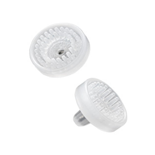
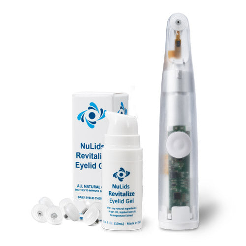
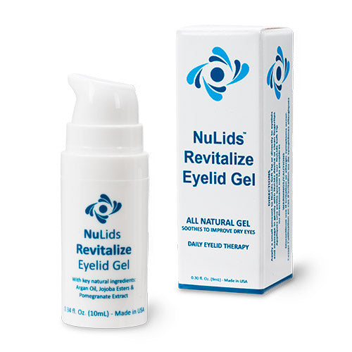
I recommend exfoliating the lid margin daily with NuLids, a recently released eyelid hygiene device that patients use at home. In addition to keeping glands open and oils flowing, NuLids use maintains a healthy level of bacterial colonization of the lids and restores ocular surface homeostasis.
Clinical improvements in blepharitis, I believe, are contingent upon daily, safe, effective, mechanical debridement. For the first time, in NuLids, we have an instrument that can fulfill that contingency.


